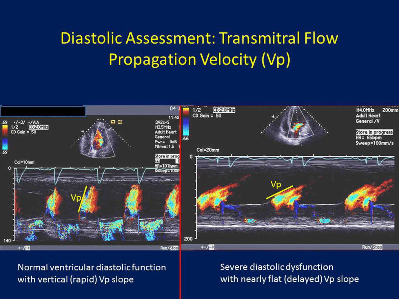
Figure 3.
Diastolic assessment: Transmitral flow propagation velocity (Vp): Placing the color Doppler sample volume from the mitral annular level to the left ventricular (LV) apex, the more rapid the LV relaxation, the faster blood travels from the mitral annular level to the LV apex, and hence the more vertical the color Doppler mitral inflow m-mode and the more rapid the Vp slope (left panel). On the other hand, the more impaired the LV relaxation, the slower it take for blood to go from the mitral annular to the LV apex, hence a “flatter” Vp slope (right panel).
© 2015 Dokainish, licensee Bloomsbury Qatar Foundation Journals.
الاقتباس:
